Medical physics
Medical physics
The following text is used only for teaching, research, scholarship, educational use and informative purpose following the fair use principles.
We thank the authors of the texts and the source web site that give us the opportunity to share their knowledge
Physics
Medical physics
What is Medical Physics?
Medical physics is the application of physics to medicine. A large part of medical physics concerns the use of radiation to examine and treat the body. Other areas include medical ultrasound, endoscopy and MRI scans.
History
Most people think that Medical Physics started in 1895 when Wilhelm Roentgen discovered x-rays but, in practice, physics has been used to investigate the body for much longer than this.
Hippocrates (460-377 BC), the “Father of Medicine”, may have been the first medical physicist. Over two thousand years ago, he wanted to know where an infection was on a patient’s back. He smeared mud over the patient’s back as he knew that infected tissue is warmer and would therefore dry the mud faster.
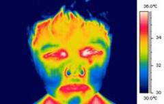 Technology has improved since then, and modern thermography, which looks at heat coming from the body using an infrared camera, is very different from Hippocrates' methods.
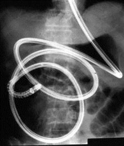
When doctors wanted to be able to see inside the stomach and intestines, they first simply used a thin tube with light provided by a candle. In 1868 a metal tube was passed down the throat into the stomach. This was an early prototype of an endoscope. Large early models were tested on sword swallowers, and were very intimidating, with one man quoted as saying 'I'll swallow a sword, but I'll be damned if I’ll swallow a trumpet'. Modern endoscopes are smaller and more flexible, making endosopy a less unpleasant examination.
In the future, we will use camera pills that can be swallowed and travel all the way through the digestive system without any discomfort.
The most common medical imaging examination today is an x-ray, or radiograph. X-rays were discovered in 1895 by Wilhelm Roentgen, who was passing an electric current through a glass tube with a vacuum inside, when he noticed a screen nearby start to glow. He realised that some invisible rays from the tube were causing the glow, and called them x-rays as he didn't know what they were. He set to work, trying to find out more about these strange rays.
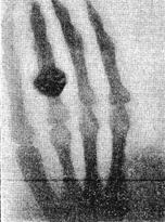
His wife became worried that he was spending so much time in his lab, and wasn't eating properly or talking to anyone. She finally persuaded him to tell her what he was working on, and he took her into the lab. She tried out his equipment by putting her hand in the x-ray beam for fifteen minutes, and saw an image of her hand appear on film behind it. This was the first medical x-ray image. Roentgen was awarded the first ever Nobel prize in Physics in 1901.
Soon after Roentgen’s discovery, Henri Becquerel, a Frenchman, was experimenting with salts that fluoresced when exposed to sunlight to see if they would emit x-rays. One cloudy day he left a photographic plate and a uranium compund in a drawer, and when he developed the film he found that it had been exposed to something, even though it hadn’t seen any light. He realised that uranium gave off invisible rays that could ionise atoms and blacken film. He called this radioactivity.
Marie Curie, along with her husband Pierre, continued the research into radioactive materials, discovering new radioactive elements Polonium (named after her homeland, Poland) and Radium. Bequerel and the Curies shared a Nobel prize for their work.
For a while radiation was hugely fashionable, with people putting radium in water and thorium in toothpaste. This health craze was dangerous though, and ill effects were soon noticed. The girls who painted radium onto the dials of watches developed thoat and mouth cancers from licking their brushes, while people using radioactive products suffered symptoms such as burns, hair loss, bone diseases and various types of cancer. Marie Curie herself died from a blood disease linked to radiation exposure (Pierre, though suffering from radiation sickness, died when run over by a horse and cart) .
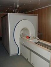
 By this time, it had been realised that, although harmful in large doses, small amounts of radiation could be used to treat diseases such as cancer. The radioisotope cobalt-60 became used in radiotherapy machines (see left) and today radiotherapy uses high energy x-ray beams to treat tumours.
Radiation Physics
Ionising radiation can be either alpha or beta particles, or high energy electromagnetic waves with enough energy to completely remove an electron from an atom.
How does radiation affect the body?
In large doses, ionising radiation can be dangerous and can cause burns to the skin and sickness. In medical imaging radiation doses are too low to cause serious damage, though there are still risks. Radiation can damage the DNA in cells. Sometime the cells can repair the damage, though sometimes they cannot and die. This fact is useful in the treatment of cancer where radiotherapy is used to kill the cells in a tumour. Another problem is that radiation can change the DNA without the cell dying. This is called a mutation, and mutations can lead to the person being more likely to develop cancer later in life, or can be passed on to their children, sometimes leading to genetic abnormalities.
X-Radiography
X-ray radiography is one of the most commonly used methods of diagnosis. It can be used to examine broken or fractured bones, teeth, the digestive system, the lungs and to detect breast cancer.
X-rays are produced when electrons hit a metal, which in hospital x-ray tubes, is usually tungsten. The x-rays then pass through the body and onto either a film cassette or digital detector (like in a digital camera).
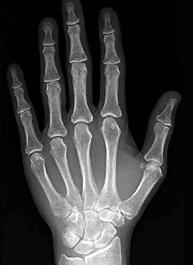
Structures in the body like bones are very dense and contain elements such as calcium that have a high atomic number. This makes bone absorb a high proportion of the x-rays. Soft tissues like fat and muscle allow more x-rays to pass though. The body casts an x-ray shadow onto the film. Where the x-rays have passed though bone, the film is less exposed so it looks white; where they have not passed though anything the film is exposed and turns black; and where the x-rays have passed through soft tissues the film has different levels of grey.
In order to make some parts of the body show up better, contrast media with a high atomic number can be used. This can be a 'barium meal', where the patient drinks a liquid containing barium (atomic number 56) which makes the digestive tract show up clearly on x-rays, or the patient can have an injection of iodine (atomic number 53) which makes the blood vessels stand out (this is called angiography).
CT (computed tomography)
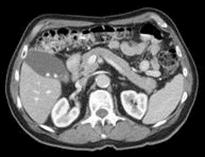
 A CT scan (sometimes called computed axial tomography, or a CAT scan) also uses x-rays.
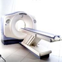
In a CT scan the patient lies on a table and is moved though a doughnut-shaped machine. It creates images that are slices through the patient.
 It does this by moving the x-ray tube and detector in a circle taking x-ray images of the slice from all angles around the body.
A computer then processes these images to produce a cross sectional image (a picture of a slice through the body).
CT scans are useful as they can show a range of very different tissue types clearly: lung tissue, bone, soft tissue and blood vessels.
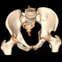
By adding together CT slices, 3-D images can be generated.
They are often used to plan radiotherapy treatments.
CT is useful for diagnosing internal inuries in trauma victims. Because a scan takes only a couple of minutes it can find problems quickly and save their lives.
One problem with x-ray CT is the radiation dose to the patient. A scan of the abdomen gives a dose of 10mSv, which is equivalent to the natural background radiation exposure over 4 years. This is about 100 times more than a standard chest x-ray.
Nuclear Medicine
Nuclear medicine uses radioactive isotopes (radioisotopes) to image the body. X-ray images show only the structure of the body, so they can be used to see things like broken bones and some tumours. Unlike x-ray images, nuclear medicine can show the function of the body. It follows what happens to certain chemicals so it can be used to see if an organ is doing its job properly. The chemicals, called tracers, are 'labelled' with a radioactive isotope and their path followed through the body.
The radioisotopes are produced in generators where isotopes with long half-lives (e.g. molybdenum-99, half-life 67 hours) decay to isotopes with shorter lives (e.g. technetium-99m, half-life 6 hours). The shorter half-lives are necessary so that the radioactivity of the patient does not remain much above its normal background level for longer than necessary.
The isotope with the shorter half life is drawn out of the generator in a solution and can be made into a range of different drugs (radiopharmaceuticals) that are absorbed by different parts of the body. The radiopharmaceutical is drawn up into a syringe shielded with lead and its dose checked before it is injected into the patient.
The gamma rays given off by the radioisotope are detected by a gamma-camera (a detector that takes images with gamma rays) which is connected to a computer and gives an image of where the isotope is in the patient. The image shows where the drug is absorbed.
If several pictures are taken over a period of time it can also show how quickly the isotope is absorbed.
These three images show the build up of a tracer in the kidneys over time. We can tell that the left kidney is blocked, as the tracer hasn’t been able to reach it.
PET (Positron Emission Tomography)
Positron Emission Tomography (PET) scanning uses beta+ emitting isotopes.
The isotope decays emitting a positron (which is a positive electron, also called a beta+ particle, and is a particle of antimatter). The positron can only travel about 1mm before losing its energy and slowing down. When it slows down enough, it will meet a negative electron from a nearby atom, and they will 'annihilate', leaving no particles. Their energy is converted into two gamma rays which travel in opposite directions so that momentum is conserved.
A PET scanner has a ring of detectors so that both gamma rays are seen, and is connected to a computer which can work out where the gamma rays came from and produces an image.
Not all hospitals have PET scanners as they need large, expensive machines called cyclotrons nearby to produce the positron-emitting isotopes. The isotopes have a shorter half-life than the gamma emitters used in traditional nuclear medicine (e.g. Carbon-11, which has a half-life of 20.5mins).
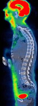 PET imaging is often used to detect tumours. As cancers are growing quickly they need a large supply of energy, which they get from glucose. A chemical called fluoxyglucose can be labelled with positron emitting fluorine-15, which then collects in the tumour and shows up as a bright spot in the PET scan (like in the rib in the picture on the right).
Some PET scanners now have a CT scanner next to them so both types of scan can be done at the same time. This can easily be done as both types of scanner are shaped.
This image is a combined PET/CT image. The excellent contrast from the PET scan, in which the brain and bladder show up as bright red, is combined with the anatomical detail from the CT (shown in grey).
Radiotherapy
Radiation is not just used for diagnosis, but for treating cancer as well. This is called radiotherapy.
Radiotherapy uses the fact that ionising radiation damages cells, and high enough doses can kill them. The cells in cancerous tissue divide very rapidly. This makes them more susceptible to damage by radiation than healthy cells, so there is a higher chance that they will be killed. Even so, care has to be taken to ensure that only the malignant cancer cells, and not the surrounding healthy tissue, receive a high dose.
This is done by mounting the system on a ring so it can rotate around the patient, with the tumour at the centre of the rotation. In this way the tumour gets a higher dose of radiation than the surrounding healthy tissue.
Originally, radiotherapy machines consisted of a cobalt-60 source which emitted gamma rays which irradiated the tumour. Modern hospitals use linear accelerators (linacs for short) instead to produce very high energy x-ray beams, with a higher energy than the Cobalt-60 gamma rays. In the UK, medical physicists are required by law to calibrate the linacs to ensure that the best possible treatment is given.
Each treatment requires careful planning. This involves deciding which directions to irradiate the tumour from, what dose to give and, in new machines, what shape region to expose.
The size, shape and location of the tumour are worked out using CT or MRI scans. Isodose curves, which join points that will receive the same dose, are drawn onto this CT scan.
Brachytherapy
In Brachytherapy (meaning short-distance therapy), radioactive material is inserted into the body, inside or near to the tumour. This means the tumour receives a high dose while the surrounding tissues have a smaller exposure.
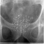
Here, tiny pellets of radioactive iodine-125 have been implanted into the prostate gland.
These pellets will not be removed, but have a fairly short radioactive half-life so that after a while they will become inactive.
The Gamma Knife
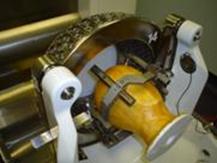
The gamma knife is not really a knife, but a way of performing brain surgery without cutting through the skin, muscle or skull. It uses 201 radioactive cobalt-60 sources to irradiate the brain. Cobalt-60 emits gamma rays and has a half-life of 5.26 years.
The first stage in treatment is to fit a metal frame to the skull, which is done using four screws under local anaesthetic. The rigid frame allows the radiotherapy to be performed very precisely.
Then, the treatment is planned using CT or MRI images, so that the sources are correctly targeted, to irradiate the tumour and avoid healthy tissue, especially sensitive regions around the eye and cochlea.
The Cobalt-60 sources are positioned in a hemisphere. The patient’s head, held in the frame, is held inside a helmet with 201 holes to precisely target the radiation. When treatment starts, the patient’s head is moved inside the unit.
The gamma knife is used to treat benign and malignant tumours, blood vessel malformations, some pain conditions and some movement and psychiatric disorders. In 2006, there were three in the UK (two in London and one in Sheffield).
Radiation Protection
Why do we use radiation?
The doses of radioactivity used in medicine are small, and the benefit of being able to find out what is wrong with a patient and then treat them often outweighs the increased risk of possibly developing cancer later in life.
We all receive a dose of radiation from background sources such as radioactive rocks, radon gas and cosmic rays. This can be between 1.5 and 7.5 mSv per year on average, depending on where you live. Compare this to the dose from a dental x-ray, which is about 0.01 mSv, the equivalant of about 1½ days background radiation.
 A chest CT scan gives a radiation dose of about 8 mSv, which is about the same as 3½ years background exposure, but you would receive the same dose from a four hour flight, about the time it takes to fly to Greece from London, as you are higher up and have less atmospheric protection from cosmic rays. This dose increases your risk of developing cancer by one in 2500, though your risk without ever having had an x-ray is already 1 in 3.
How can we protect ourselves?
There are many ways to reduce the dangers from radiation. The first is only to use it when necessary. Before people realised it was dangerous, shoe shops used to x-ray people’s feet to check that new shoes fitted properly. This no longer happens as the benefit did not justify the risk. However, the benefits of seeing where a bone is broken so it can be safely and properly mended are considered worth the small extra risk.
Every x-ray examination has a strict controls about the maximum radiation dose a patient can be given, and the patient can be covered with lead-rubber shield to protect the parts of them not being examined from the radiation. This is especially used to protect reproductive organs so there is less risk of a mutation being passed on.
People such as radiographers and nurses who work with radiation every day will leave the room or stand behind a lead shield when a procedure takes place, as the risk of developing problems due to radiation exposure increases with total dose. They also wear film badges which are developed regularly to check the dose they have recieved.
What jobs do medical physicists do?
Medical physicists work in many areas of the hospital. They are often responsible for maintaining and calibrating sophisticated equipment like MRI scanners and linear accelerators. They may develop new ways of using equipment to diagnose and treat unusual conditions. They may also be responsible for hospital computer systems. In general, they ensure that all the high-tech equipment needed in a modern hospital is working well so patients can be diagnosed safely and quickly.
Many medical physicists also work in research in universities, industry and hospitals, improving existing techniques and developing completely new ones. People such as Sir Godfrey Hounsfield and Sir Peter Mansfield have won Nobel prizes for their work in CT and MRI respectively.
Who does medical physics?
Roughly half of medical physicists are women and half are men. If you’re considering a career in medicine, physics or engineering, then medical physics may appeal to you.
Medical physicists use a wide range of skills including writing computer programs, inventing new equipment, and working with patients.
Ultrasound
Ultrasound imaging
Ultrasound uses sound waves with frequencies between 1 and 10MHz to look inside the body. These frequencies are too high to be heard by humans. The ultrasound waves, like all waves, can be reflected, refracted or transmitted at boundaries. It is the reflections, or echoes, which are used to produce ultrasound images.
A gel is used so the probe makes good contact with the skin. It sends out pulses of ultrasound, and measures the time taken to detect the echo and the strength of the signal. The time taken indicates how deep in the tissue the ultrasound wave is being reflected.
Ultrasound imaging is particularly good at detecting cysts, which are pockets of fluid, in the liver, glands and ovaries and breasts, and can be used to identify gallstones and kidney stones, which are deposits of minerals. Large blood vessels also show up clearly.
Ultrasound is commonly used during pregnancy to check the development of the foetus. It can show the size of the foetus which indicates how far along the pregnancy is, check that the heart is beating and identify problems.
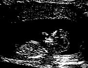
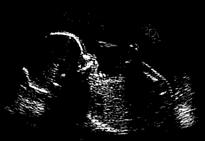
It is thought to be safe as it doesn’t use ionising radiation.
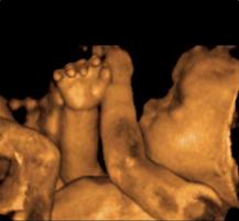 Computers can now generate 3-D ultrasound images, and 4-D (3-D over time) ultrasound scans can be made into videos for parents.
This image is processed to show the skin. This is called surface rendering.
Doppler Ultrasound
The Doppler effect is the change in the frequency of a sound due to the person listening moving relative to the source of the sound. If you move towards a source (or stand still and it moves towards you) the pitch, or frequency of the sound, increases. Likewise, if you move away from the source, the pitch of the sound decreases and it sounds lower. This can be easily noticed in the pitch of an ambulance siren as it gets closer, passes you and then moves away.
The Doppler effect is used in medicine to study blood flow. It can tell you if blood is moving towards or away from the probe.
This is a Doppler ultasound probe. It is being used to examine blood flow in the radial artery, the same one that you would use to measure your pulse.
probe
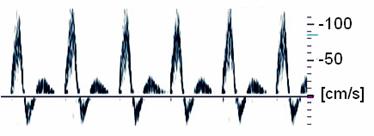
This is the Doppler ultrasound signal measured from a healthy artery. When the heart beats it pushes blood at a velocity of over 100cm/s away from the probe.
The information can be colour coded and combined with conventional ultrasound images, which is particularly useful in diagnosing blockages in blood vessels.
This combined image shows the blood is all flowing in the same direction. This indicates that the blood vessel is healthy.
In this image some blood is moving away from the probe and some blood towards it. This is turbulent flow, like rapids in a river, and is caused by a blockage in the blood vessel.
Optical methods
Endoscopy
 Endoscopy is a way of looking inside the human body through a narrow, flexible scope. It is mostly used to diagnose problems in the oesophagus, stomach and intestines, including ulcers, bleeding and tumours. If something suspicious is seen, a biopsy (a small sample of tissue) can be taken and examined later by a pathologist to see what it is. Typically optical fibres are used to transfer light to the end of the endoscope and a miniature video camera records the image.They also have a biopsy channel (along which tissue can be taken or other surgical instruments can be passed) and water pipe for washing the field of view clear.
Laparoscopy is an extension of this technique where the scope is used to look inside the abdomen and pelvis through a small cut, or incision.
In endscopic surgery, commonly known as keyhole surgery, the endoscope is passed through an incision into the patient and the surgeon, who uses knives or lasers also passed though the scope, watches what he is doing on a video screen. Keyhole surgery can be used to treat hernias and remove tumours and is often used on sportstars' injuries as the recovery time is faster than in normal open surgery.
Endoscopes for remote robotic surgery are currently being tested. In 2001, doctors in New York removed the gall bladder of a woman in France using an endoscope remotely. Endoscopic pills which include a camera and transmitter are currently being developed. This would allow the whole digestive tract to be examined painlessly.
Pulse Oximetry
Oxygenated and deoxygenated blood are slightly different colours and so absorb different frequencies of light differently. By looking at the absorption of two different frequencies of light, we can distinguish between blood carrying oxygen and blood not carrying oxygen.
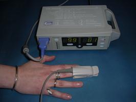
This principle is the basis of one of the most commonly used instruments for monitoring the body - the pulse oximeter. A pulse oximeter clips onto a finger (or a baby's foot) and has inside it one red light source, one near-infrared light source and a detector. As the blood pulses, more blood enters the finger and the amount of light detected decreases. However, the decrease in the amount of red light differs from the decrease in the amount of near-infrared light. The size of this difference depends on the amount of oxygen in the blood. At the same time, the blood moves through the finger in pulses, allowing the heart rate to be measured.
The pulse oximeter is often used to monitor the well being of patients in intensive care and anaesthetised patient.
Laser Surgery
Lasers produce light that has only one wavelength, rather than a range of wavelengths like most other light sources. They are very useful in surgery as they can be focussed to a small point, enabling them to vaporise, seal or cut tissue.
Eye surgery
Laser eye surgery can be used to correct long or short sight, and astigmatism (distorted vision). A surgeon cuts a thin layer of the cornea off to create a flap. A laser is then used to cut and reshape the cornea behind the flap. The flap is then closed and grows back naturally.
Hair removal
When laser light is shone onto the surface of the skin it is absorbed by melanin, the pigment that gives hair and skin their colour, and is converted into heat. If enough energy is absorbed, the part of the hair follicle that causes hair growth is destroyed, and the hair cannot grow back.
As the skin contains melanin as well, it also heats up and can be damaged.
Two things allow laser hair removal to be done safely:
- the hair follicle contains more melanin than the skin
- the surface area of skin is larger so it cools down faster than the hair follicle
Laser hair removal therefore works best on people with pale skin and dark hair. A tan is caused by extra melanin being produced, so you should wait for a tan to fade before having laser treatment.
Mole Removal
Mole removal works in a similar way to hair removal.The pigment in the mole absorbs the energy from the laser light and is broken up. It is then carried away by the body and when the skin heals its colour is the same as the surrounding skin.
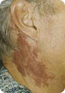 Port Wine Stains
Blood vessels can widen (dilate) to allow more blood through, so when you exercise and your body needs to get rid of heat, the blood vessels in your skin dilate and you look red.
![]()
A port wine stain is an area of red or purple skin. They are caused by blood vessels always being dilated, so the skin always looks red as it permanently contains a lot of blood. About 3 in 1000 babies are born with a port wine stain.
Lasers can be used to destroy the tiny, dilated blood vessels, without harming the surrounding skin. Treatment works better on children than on adults. 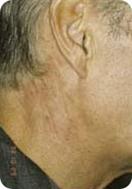
Magnetic Resonance Imaging (MRI)
MRI is a way of looking inside the body and is especially good at producing images of soft tissues such as muscle, fat, cartilage and the brain. It does this by producing a map which depends on the density of hydrogen in the body.
MRI uses a very strong superconducting magnet with a magnetic field strength of around 40 000 times that of the Earth. The nucleus of a hydrogen atom is a single proton, and is like a little bar magnet.
![]()
 When a person is lying in the magnetic field of the MRI scanner the nuclei of the hydrogen atoms in their body line up, like compass needles in the Earth's magnetic field, either pointing in the direction of the field or opposite to it.
 The hydrogen nuclei (protons) don’t stay still though, but move like a spinning top around the direction of the magnetic field.
A radiofrequency field, an alternating magnetic field that has the same frequency as radio waves, is then applied. This flips some of the protons round and makes them all move round together. This produces a changing magnetic field at right angles to the large magnetic field, which can induce a voltage in a coil of wire. This signal can be used to produce an image which, which depends on the number of protons and how tightly they are held by surrounding molecules.
A third magnetic field has a gradient so it is stronger at one end than the other. This allows the scanner to select a slice of the body to look at, by selecting the required field strength. The gradient fields change rapidly and make the scanner very noisy.
 MRI is used for diagnosing many problems. It can be used to identify tumours, diagnose multiple sclerosis (MS) and is often used on sportspeople to see problems with ligaments inside joints like the knee and ankle. It can also be used to show the anatomy of the brain and how it works.
 MRI doesn't use radiation, and magnetic fields are thought to be safe. However, MRI scanners are very big and expensive. Also, because of the strong magnetic fields, all metal objects have to be kept out of the room or they would get pulled into the scanner. People with pacemakers or other implanted devices can't have MRI scans as the magnetic field would stop them working. An MRI scan can take up to 20 minutes to complete and you have to be still the whole time as any movement would blur the image. The changing magnetic fields also produce a lot of noise, which can be scary, and as you are inside the scanner during the scan, people with claustrophobia can find the process upsetting.
MRI produces images that are 2‑D slices through the body and they have excellent spatial resolution (i.e. you can see very small details in the images), making it an important tool for doctors.
ECG (Electrocardiogram)
If a patient complains of chest pains or shortness of breath it is important to check that their heart is working properly. The heart is a muscle made up of four chambers which contract to push blood and around the body. The messages that tell the muscle to contract are electrical signals, and by measuring these signals we can see if there is anything wrong with the heart. This is done using an ECG.
To take an ECG, three electrodes are placed on the surface of the skin: one on the right arm, one on the left arm, and one on the left leg. These contacts are connected to a machine called an electrocardiograph. This draws lines on graph paper showing the electric potential between each electrode over time.
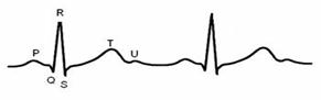
Each voltage spike is due to an electrical signal in the heart and is represented by a letter. A healthy heart should give a trace that looks something like this.
Changes to this pattern could indicate a problem such as missed beats, no P-wave (the atria not contracting) or very high spikes (the ventricles working too hard due to high blood pressure).
Bioengineering
Pacemakers
The muscles of the heart contract in a regular rhythm and with a steady pace of about 70 beats per minute for someone at rest, and higher for someone who is exercising. The electrical signals that tell the heart muscles to contract can stop working properly, causing the heart to beat to irregularly or not at all. A pacemaker is an electronic device that makes the heart muscles contract at a regular rate.
There are two parts to a pacemaker; the generator and the leads.
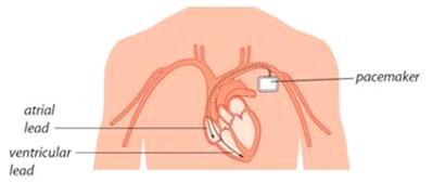
The generator is about the size of a matchbox, and contains the battery and a mini computer that processes information about the electrical signals in the heart and sends out its own signals to tell the heart to beat. It is implanted just under the skin by the left shoulder. The batteries last for up to ten years, and are checked regularly.
The leads are passed into the heart though a vein coming from the left shoulder, and are implanted in the muscle of the wall of the right atrium and ventricle. When the generator sends out an electrical signal, the leads pass it on to the heart and the muscle contracts, causing blood to be pushed through and out of the heart.
Medical Engineering
Medical Engineering is the design and manufacture of all health related products. These include tools (including robotics) for surgery, scanners and hospital equipment, prosthetics or products that help people with health and disability problems live more normal, comfortable lives.

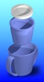
Some new products include a mug that only releases one sip at a time so people with motor neurone disease can drink more easily, portable raised toilet seats to allow people with disabilities to use public toilets, bikes for children with growth problems so they can play like other children and a 'Smart House' to allow people with dementia to live safely in their own home with as few noticeable changes as possible.
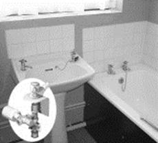 The Smart House gives verbal warnings if taps are left running, it will reduce water flow in the taps if the bath gets too full and automatically turn off the cooker if a pan boils dry or gets too hot. There are also verbal warnings by the doors saying things such as 'don't forget your keys' and sensors under the bed to automatically turn on the light if the person gets out of bed during the night.
Cochlear Implants
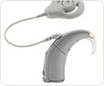
Cochlear implants are devices used to stimulate hearing in people with severe to profound hearing loss.
 |
|

They work using a microphone, processor and transmitter outside the ear (left), and a reciever and electrodes inside the head (right).
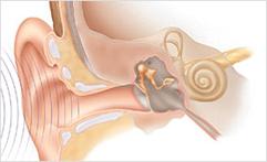 Sound waves cause the eardrum to vibrate, which sets the bones in the inner ear in motion, which in turn causes the fluid in the inner ear to move.
The cochlea in a healthy ear contains hair cells. The motion of the fluid causes the hair cells to vibrate. Nerve cells detect this vibration and send a signal to the brain. High pitched sounds which have high frequencies can only travel a short distance along the membrane; low pitched sounds can travel further. In this way the brain can tell the difference between high notes and low notes. Louder sounds don't affect the distance along the membrane they can travel, though they make the hairs vibrate more.
Damage to these hair cells, due to genetic muations, illness, chemical reactions or excessively loud noises, can lead to hearing loss.
When hearing using a cochlear implant:
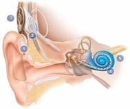 1) A microphone turns the sound into an electrical signal
2) A processor sorts the signal into groups of frequencies.
3) A transmitter sends this information to the receiver inside the head using radiowaves and the receiver sends the required electrical currents to the correct electrodes.
4) Electrodes, which are implanted inside the cochlea, stimulate the nerve cells.
5) The auditory nerve sends a message to the brain.
These devices, though they have been show to work well, are controversial. People who use sign language to communicate often see themselves as part of a community. Some people object to implants as they feel they are destroying this community.
Cochlear implants work best when implanted into young children. However, at this age children are too young to make the decision for themselves, and their parents must make the choice for them.
Supporters of cochlear implants believe that people are meant to be able to hear and an implant is therefore no different to giving a child glasses to help them see.
What do you think? ![]()
Images courtesy of Cochlear Europe Ltd.
The Future
Medical physics continues to play a valuable role in healthcare. More and more hospitals are buying MRI and PET scanners and more people with disabilities are receiving increasingly sophisticated devices.
Medical physicists are at the forefront of developments in healthcare. In the NHS, medical physicists plan radiotherapy treatments and ensure that the equipment is safe. They develop new methods using MRI and ultrasound, and assist people with disabilities. Medical physicists also work in both universities and industry, where they lead research into improving existing techniques and developing completely new ones.
For more details about jobs in medical physics, see www.ipem.ac.uk,
www.nhscareers.nhs.uk/nhs-knowledge_base/data/4847.html and
www.connexions-direct.com/jobs4u/jobfamily/healthcare/medicalphysicist.cfm?fd=1252.
For more on medical physics in general, see
www.teachingmedicalphysics.org.uk
www.ipem.ac.uk
www.cochlear.com/europe
www.gehealthcare.com
And for some medical physics games, try
www.insidestory.iop.org
Glossary
Biopsy obtaining and examining a sample of cells or tissue from the body.
Cyclotron a machine used to accelerate charged particles. For PET, protons are accelerated, then smashed into atoms which then become radioactive, emitting positrons.
Cyst pocket of tissue filled with fluid.
Diagnosis finding out what is wrong with a patient.
Endoscopy examining the body using a narrow tube.
Half-life the time taken for the activity of a radioisotope to drop by half.
Hearing loss people with profound hearing loss mostly rely on lip reading or sign language. People with severe hearing loss get help from powerful hearing aids, though may also rely on lip reading.
Ionising radiation An alpha or beta particle, or a high energy electromagnetic wave, which has enough energy to completely remove an electron from an atom.
Mutation a change to genetic material (DNA).
Near infrared electromagnetic waves that are in the infrared region of the spectrum, but have frequencies near those of visible red light (around 800 nm).
Oesophagus part of the digestive system (food pipe/gullet).
Palliative a treatment intended to ease pain and suffering rather than cure the patient.
Prosthetic something which artificially replaces a part of the body.
Radioisotope an isotope that is unstable and will decay, emitting radiation.
Superconducting A magnet made by a current flowing in
magnet a coil of superconducting wire, which is so cold it no longer has any resistance.
Therapy treating the patient.
Acronyms
CAT - Computed Axial Tomography
CT - Computed Tomography
ECG - Electrocardiogram
MRI - Magnetic Resonance Imaging
mSv - millisievert, a unit of radiation exposure
PET - Positron Emission Tomography
RF - Radiofrequency
2-D - Two dimensional
3-D - Three dimensional
4-D - Four dimensional (3 spatial dimensions and time)
Acknowledgements
This booklet was written by Emily Cook and Adam Gibson.
Many thanks to all those who contributed:
Nicola Hannam.
Jeff Jones.
Dr David Sang.
Prof. Angela Newing.
Lucy Gibson.
Dr Michael Hillman, Bath Institute of Medical Engineering.
Dr S E Jones, Gloucestershire Royal Hospital.
The Charlotte Observer Collection, Public Library of Charlotte & Mecklenburg County.
Cochlear Europe Ltd.TM
GE Healthcare Ltd.
Lumenis Ltd.
Source : http://schools.medphys.ucl.ac.uk/books_etc/textbook_a5.doc
Web site link: http://schools.medphys.ucl.ac.uk
Authors : Emily Cook and Adam Gibson
If you are the author of the text above and you not agree to share your knowledge for teaching, research, scholarship (for fair use as indicated in the United States copyrigh low) please send us an e-mail and we will remove your text quickly.
Medical physics
Medical physics
Medical physics
This is the right place where find the answers to your questions like :
Who ? What ? When ? Where ? Why ? Which ? How ? What does Medical physics mean ? Which is the meaning of Medical physics?
Medical physics physics notes
Alanpedia.com from 1998 year by year new sites and innovations
Main page - Disclaimer - Contact us